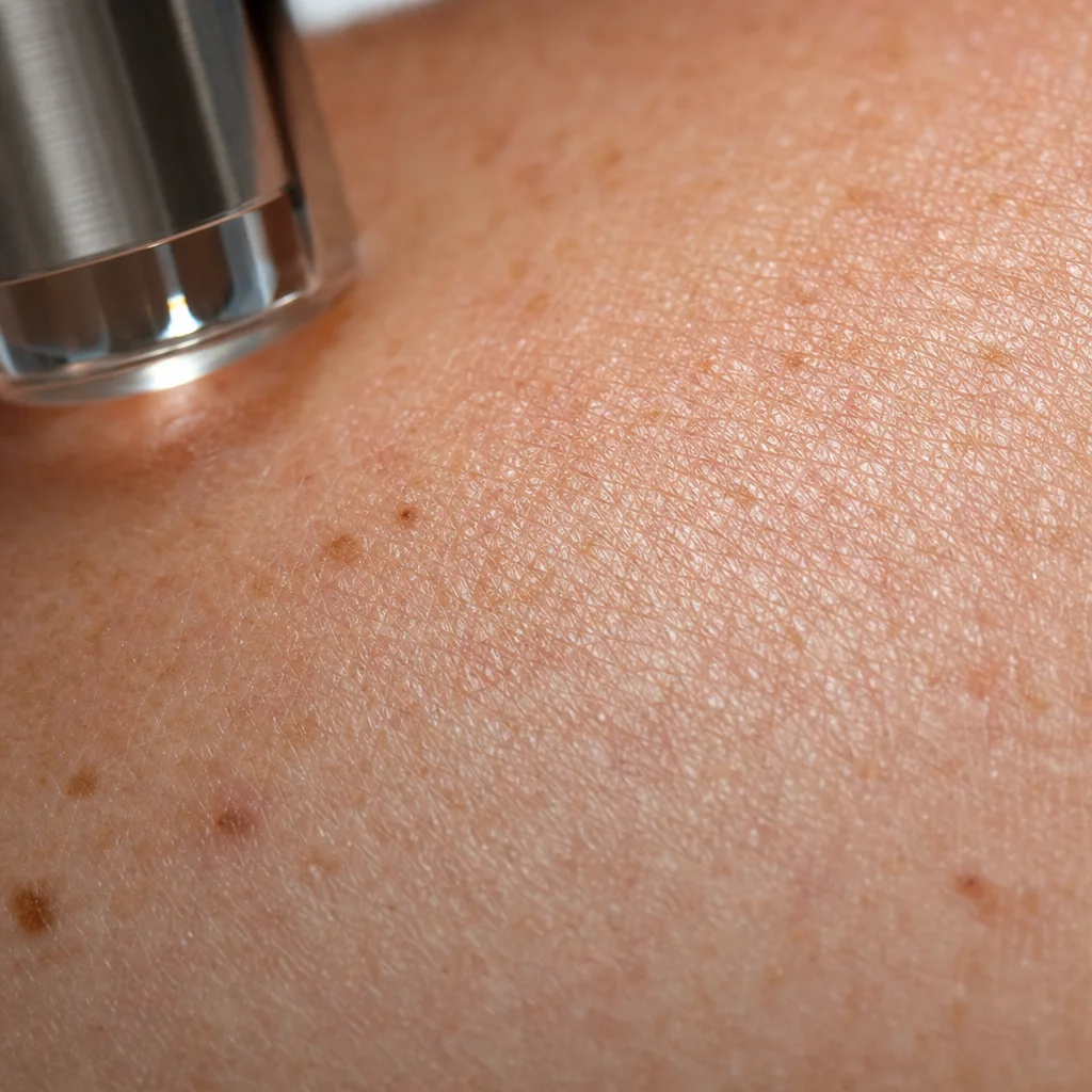
Every laser tattoo removal session is, in biological terms, a controlled wound. The deliberate delivery of intense photonic energy to the dermis triggers a cascade of cellular and molecular events that parallel the wound healing response observed in other dermal injuries — with some distinctive features unique to the photo-mechanical tissue disruption that lasers produce. Understanding this process helps explain why skin heals variably across patients, why post-treatment care matters so much, and why rushing the inter-session interval undermines results.
The wound healing response is classically divided into four overlapping phases: hemostasis, inflammation, proliferation, and remodeling. Immediately following laser irradiation, tissue damage triggers hemostasis — the arrest of any microbleeding from disrupted capillaries. Platelet aggregation and the coagulation cascade are activated within seconds. Simultaneously, the photomechanical disruption of ink particles generates a burst of damage-associated molecular patterns (DAMPs) that act as endogenous danger signals to the immune system, recruiting innate immune effectors.
Within hours of treatment, the inflammatory phase dominates. Mast cells in the dermis degranulate, releasing histamine (responsible for the immediate post-treatment edema and flare), prostaglandins, and cytokines including TNF-α, IL-1β, and IL-6. These signals drive neutrophil infiltration into the treatment zone within the first 24–48 hours — neutrophils acting as the first line of cellular defense, phagocytosing debris and generating reactive oxygen species (ROS) to neutralize any perceived infectious threat. While beneficial for clearing debris, excess ROS generation is also a key driver of post-inflammatory hyperpigmentation in melanocyte-rich skin, as ROS directly stimulates melanin synthesis through tyrosinase pathway activation.
The transition to the proliferative phase — beginning approximately 48–72 hours post-treatment — is mediated by macrophage polarization. Initially, M1-polarized macrophages continue the inflammatory response, phagocytosing ink particles and cellular debris. The critical transition to M2-polarized macrophages shifts the tissue environment from pro-inflammatory to pro-healing. M2 macrophages release growth factors including TGF-β, PDGF, and FGF-2, which stimulate fibroblast migration into the wound zone and initiate de novo collagen synthesis. This phase is why the skin may appear somewhat firm or slightly raised for weeks after treatment — new collagen matrix is being deposited.
The epidermal repair component of post-laser healing is particularly important given that even non-ablative tattoo removal lasers produce transient disruption of epidermal barrier integrity. Keratinocyte proliferation at the wound margin begins within 24 hours, driven by EGF receptor signaling. Complete epidermal regeneration over a treated area typically requires 5–14 days depending on treatment intensity and patient healing capacity. During this window, the compromised barrier significantly increases transepidermal water loss (TEWL), making adequate moisturization and occlusion an evidence-based — not merely cosmetic — aftercare recommendation.
The remodeling phase, which can persist for 6–18 months after a single aggressive session, involves the replacement of provisional type III collagen with mature type I collagen, mediated by matrix metalloproteinases (MMPs). The net effect on skin texture and appearance depends on the balance between collagen production and degradation. When post-treatment inflammation is excessive or prolonged — as occurs with inadequate sun protection, early re-treatment, or secondary infection — disordered collagen deposition can produce hypertrophic scarring. Conversely, when inflammation is appropriately modulated and healing conditions are optimized, remodeling can sometimes improve skin texture over baseline, explaining the anecdotally observed skin quality improvements some patients report after complete tattoo removal.
From a translational research perspective, the identification of molecular targets to accelerate healing and reduce PIH has generated several investigational strategies. Topical niacinamide, a well-established melanogenesis inhibitor, has been evaluated in two small randomized studies with promising results for PIH prevention. Low-level light therapy (LLLT) using 630–850 nm wavelengths has shown capacity to accelerate fibroblast proliferation and reduce post-procedural erythema duration in randomized trials. Oral antioxidant supplementation (vitamin C, vitamin E, polypodium leucotomos extract) has theoretical support from ROS-reduction data, though large clinical trial evidence for meaningful clinical benefit in the laser wound healing context remains limited.
The science of laser wound healing continues to evolve rapidly, with single-cell RNA sequencing approaches now providing unprecedented resolution of the cellular dynamics within treated skin. These tools are expected to identify specific cell populations and molecular checkpoints whose modulation could dramatically improve healing outcomes — particularly for high-risk patients including those with darker skin phototypes, keloid predisposition, or immunosuppression.