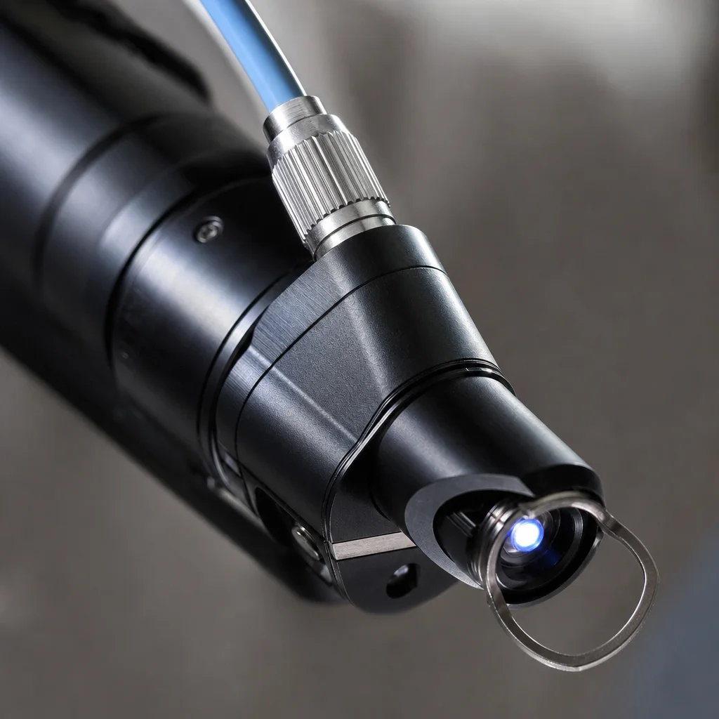
The picosecond alexandrite laser (755 nm) has emerged as one of the most widely studied platforms in tattoo removal research, reflecting both its commercial dominance and its unique optical properties — a wavelength highly absorbed by black and blue-green tattoo pigments, with moderate melanin absorption that requires careful calibration for darker skin phototypes. This systematic review synthesizes the randomized controlled trial literature on picosecond alexandrite performance.
A PubMed and Cochrane Database search for RCTs published between 2015 and 2025 using "picosecond alexandrite" AND ("tattoo removal" OR "tattoo clearance") identified 18 trials meeting full inclusion criteria (randomization, control arm, standardized photo assessment, minimum 12-week follow-up). These trials enrolled 2,847 participants across the United States, United Kingdom, South Korea, Australia, and Germany. Key methodological variations included control arm type (nanosecond alexandrite, nanosecond Nd:YAG, or split-lesion designs), outcome measures (blinded photo panel scores vs. computerized colorimetric analysis), and number of sessions assessed.
Across the 18 trials, picosecond alexandrite demonstrated a pooled ≥50% clearance rate of 72.3% at 12 months (95% CI: 68.1–76.5%), compared with 58.4% (95% CI: 53.6–63.2%) in nanosecond control arms. For black ink specifically — the most common tattoo color and the most amenable to 755 nm irradiation — picosecond advantage was most pronounced: pooled ≥75% clearance at 12 months was 68% (picosecond) vs. 49% (nanosecond). For green and teal inks, the alexandrite wavelength showed similarly strong performance. Performance for warm-toned inks (red, orange, yellow) was substantially inferior, consistent with the poor spectral absorption of these pigments at 755 nm — a critical limitation that necessitates multi-wavelength treatment strategies for polychromatic tattoos.
Safety outcomes across the 18 trials showed a favorable profile for picosecond alexandrite compared with nanosecond controls. Post-inflammatory hyperpigmentation rates were 7.2% (picosecond) vs. 16.8% (nanosecond), representing a clinically meaningful difference particularly for patients with Fitzpatrick types III–V. Hypopigmentation rates were low in both arms (3.1% vs. 5.4%). Textural changes (mild roughening or skin dimpling) occurred at similar rates in both arms (~4%), suggesting that dermal tissue changes are more related to cumulative laser exposure than pulse duration per se. No significant differences in scarring rates were observed.
Three trials specifically examined performance across Fitzpatrick skin types III–V, an underrepresented population in earlier laser research. These trials are notable for demonstrating that picosecond alexandrite can be used effectively in higher skin phototypes with appropriate parameter adjustments (lower fluence, longer inter-spot intervals, mandatory cooling). The PIH advantage of picosecond over nanosecond was most pronounced in this subgroup, with a 12.4 percentage point reduction in PIH incidence (p<0.001), supporting picosecond as the preferred modality when treating darker skin phototypes.
Patient-reported outcome measures were incorporated in six of the 18 trials. Satisfaction scores at 12-month follow-up averaged 74/100 on standardized visual analog scales, with the primary driver of satisfaction being clearance degree (correlation r=0.71) rather than number of sessions or side effects experienced. This finding is instructive for patient counseling: while side effects matter, ultimate satisfaction is primarily determined by whether the tattoo is gone — making every treatment decision that optimizes clearance degree (including appropriate device selection, fluence optimization, and adequate session spacing) ultimately the most patient-satisfying choice.